PCV
Packed cell volume (PCV) or haematocrit...
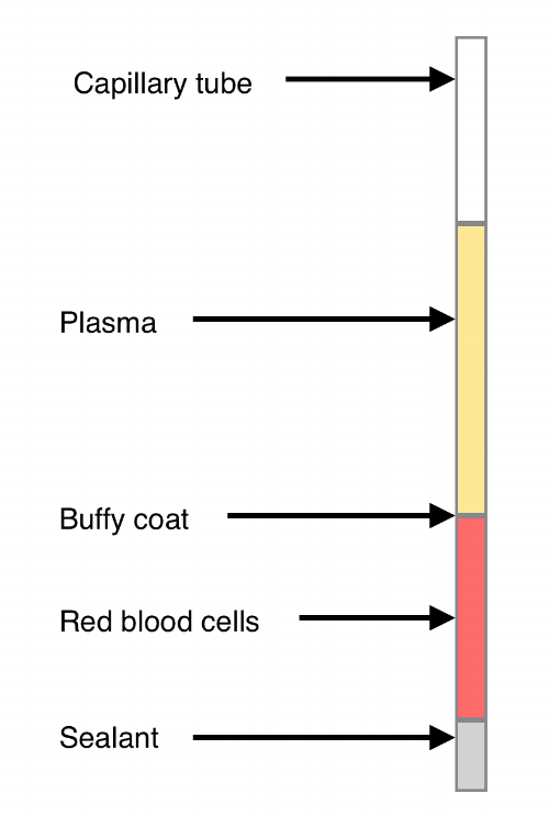
Layers in a centrifuged blood sample
This test measures the proportion of red blood cells/corpuscles (RBCs) in a centrifuged blood sample. It is useful in the diagnosis and management of anaemia, dehydration and other conditions.
Equipment:
Anticoagulated whole blood (normally in EDTA)
Clay sealer
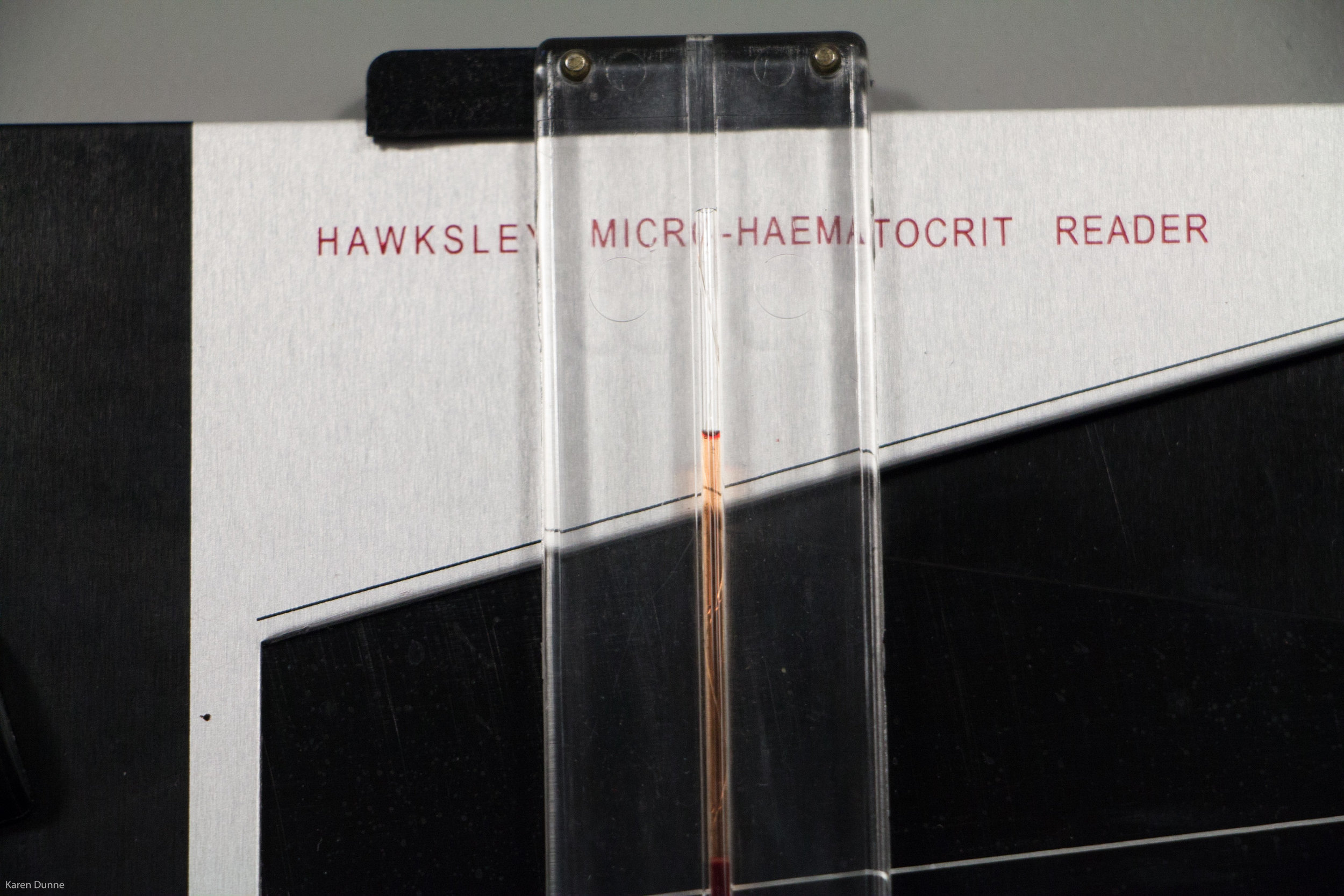
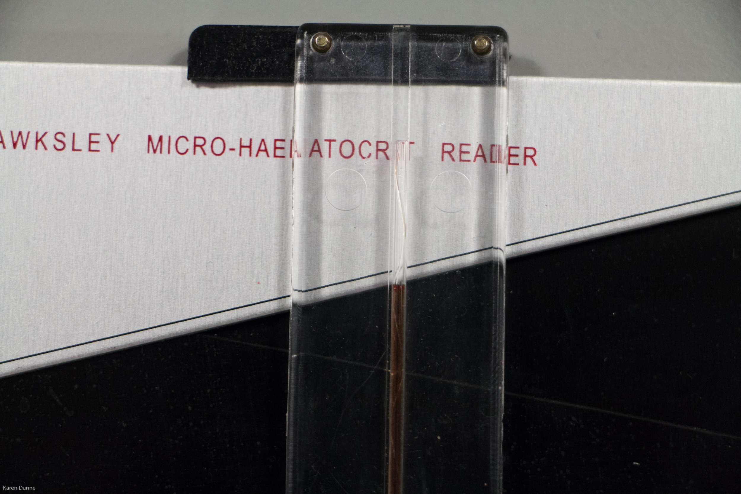
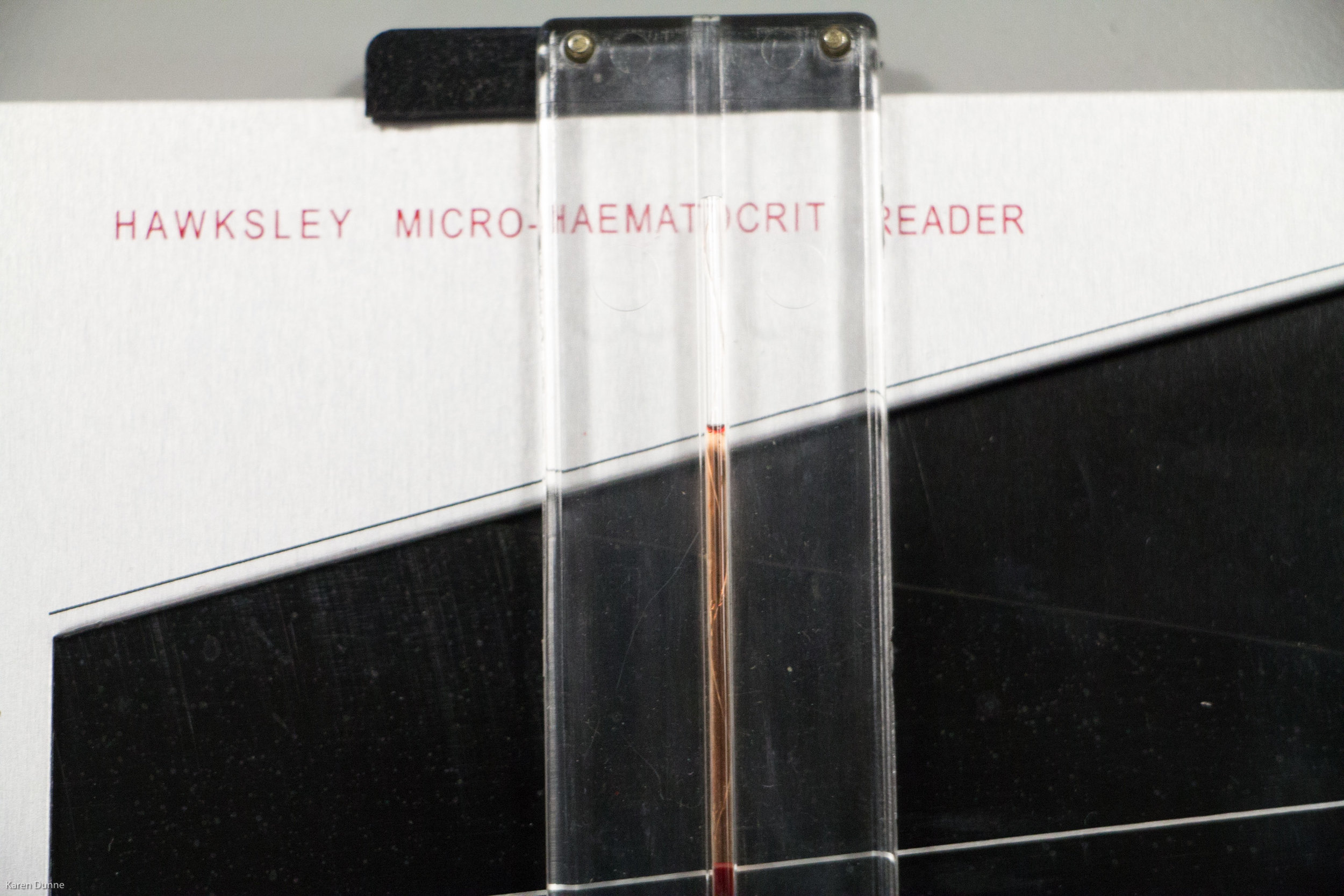
PCV reader (card or Hawksley model)
Centrifuge
Capillary tubes
Disposable gloves
Tissue paper
Sharps container
Clinical waste bin
Refuse bin
Don gloves onto clean and dry hands. Mix the sample thoroughly by inverting it gently at least five times. Remove two capillary tubes from the container. Open the blood sample and place the capillary tubes in it. Tilt the sample to about 45 degrees to assist filling of the tubes.
Once the tubes are about 2/3 full, place your finger over the open end before removing them from the blood sample (this helps to prevent air bubbles forming in the other end of the tube). Keep your finger over the end of the tubes while wiping their exterior free of blood and sealing the ends that were placed in the sample by pressing them into the clay. Once the tubes are sealed you can take your finger off the other end.
Place both tubes in the centrifuge grooves directly opposite one another, this ensures the centrifuge is balanced. Ensure the clay seals are towards the outside. Screw the centrifuge cover into place and close the lid. Spin at 10,000rpm for five minutes (the speed and time may vary on some models).
Balanced centrifuge
Each capillary tube has one opposite it (note: the lid has yet to be screwed into place before the machine is turned on).
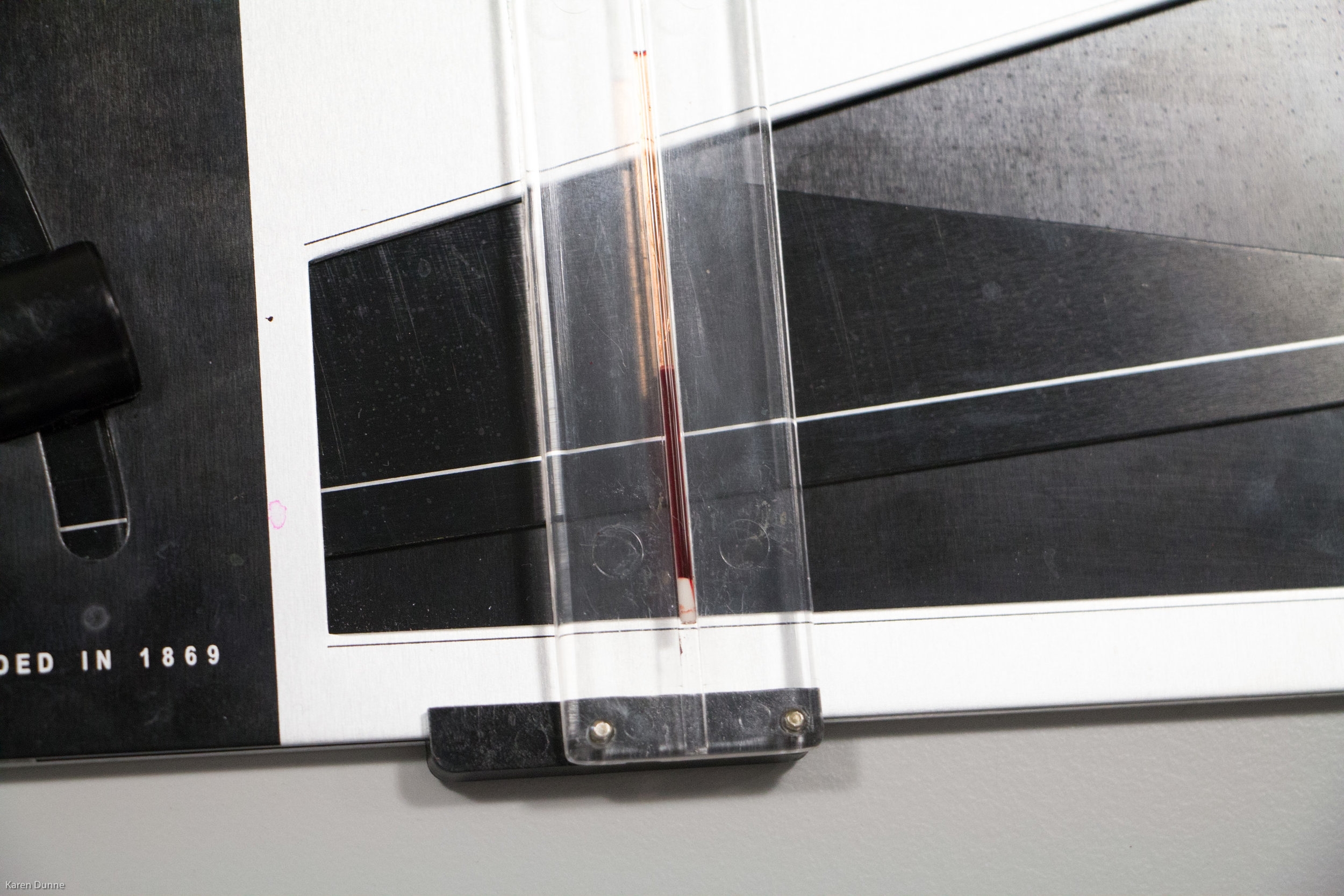
Once the centrifuge has completed its spin remove a capillary tube and examine it visually. Normal plasma is a pale clear yellow liquid. A pink or red tinge indicates haemolysis, dark yellow/orange = jaundice and milky while = hyperlipidaemia. Note also the proportion of the sample that consists of the RBCs. 30-40% is typical but this may be reduced in cases of anaemia or increased in dehydrated animals. Check for the the buffy coat to confirm the presence of WBCs in the sample (it may be absent in cases of severe leukopaenia). Buffy coat smears have been used to diagnose mast cell tumours and leishmania.
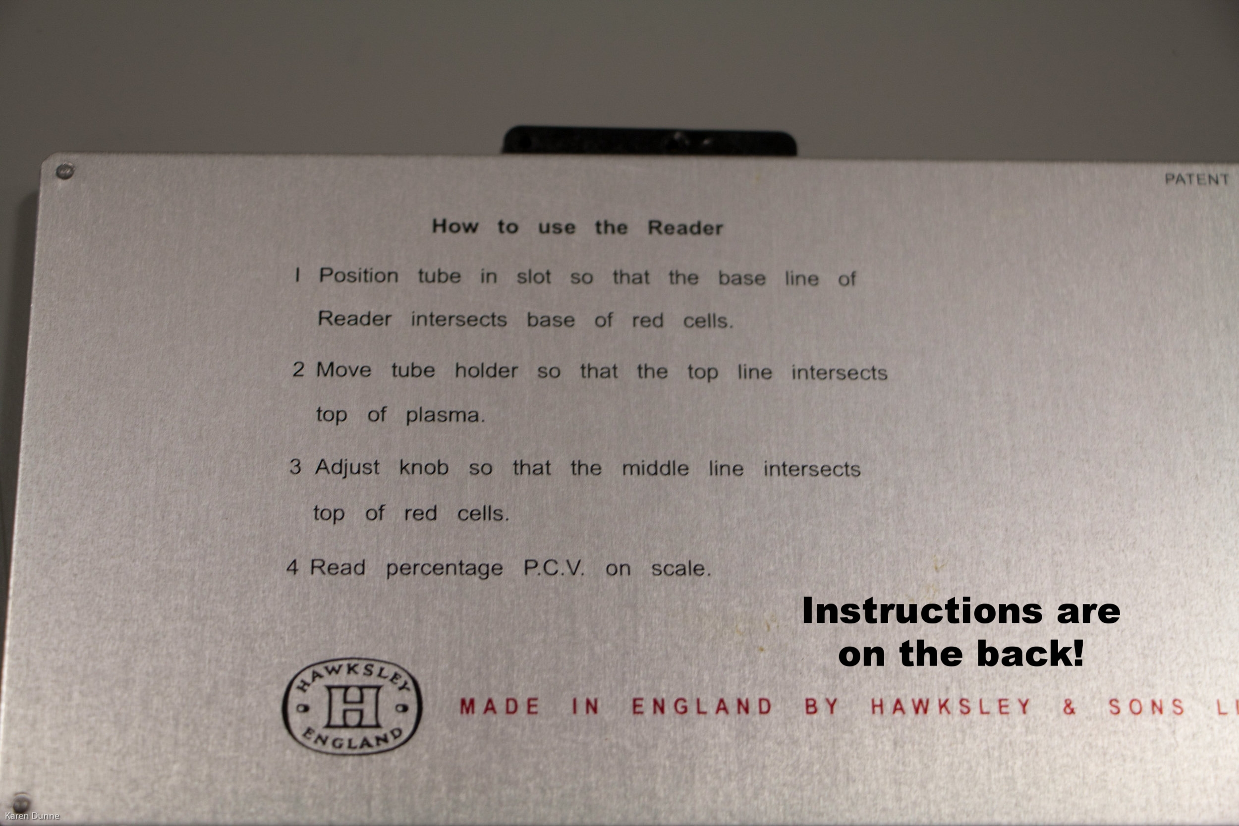
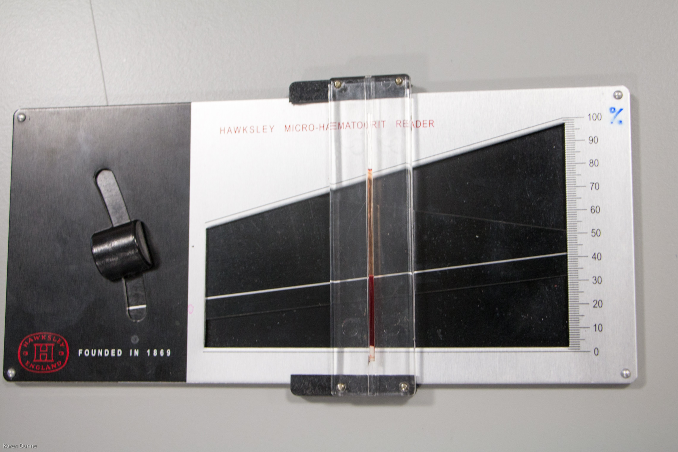
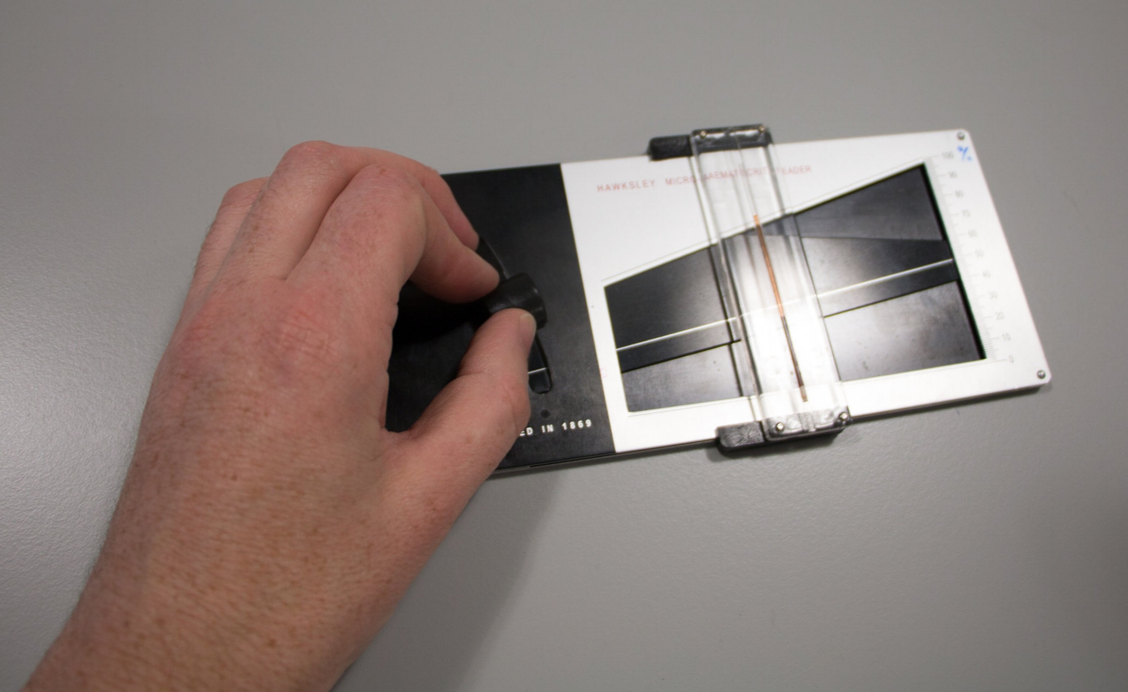
To obtain the PCV reading place the capillary tube either onto a reader card or into the tube holder on a Hawksley haematocrit reader (see slideshow below). Align the bottom of the RBC column with the 0% line and the top of the plasma column with the 100% line. The measurement is taken at the top of the RBC column and is epxressed as a percentage (%). Repeat for the second tube. For quality control purposes each reading should be within 1% of the average.