
Diagnostic Imaging: equine limb radiography

Veterinary nurses need to be able to safely produce high quality diagnostic images
Equine distal limb radiography
It's essential that you know your anatomy so that you obtain a diagnostic image of the correct site without exposing yourself, your colleagues, your clients or your patients to unnecessary radiation!
Revise structure and function of the horse's forelimbs with Dr. Roberta Dwyer of the University of Kentucky's Gluck Equine Research Center.
Revise structure and function of the horse's hind limbs with Dr. Roberta Dwyer of the University of Kentucky's Gluck Equine Research Center.
Equine radiography overview
Taking x-rays of a horse is normally a three person task:
- One person to hold the horse
- One person to hold the cassette
- One person to operate the machine and make the exposure
Sometimes you don't need a person to hold the cassette e.g. if using a block to obtain a foot x-ray.
Patient preparation
The area to be imaged must be clean and dry - brush off dust, mud and loose hair using a suitable brush e.g. a dandy brush for dried mud, a body brush on sensitive skin areas (revise equine grooming!). If obtaining foot images the shoe should be removed (revise shoe removal) and the hoof meticulously picked out and scrubbed clean using a stiff brush (dandy brush is suitable). Some people like to pack the solar sulci with modelling clay to avoid air shadows around the frog.
All personnel must be at least 18 years old, non-pregnant and used to handling horses. The horse will need a head collar and lead rope in all cases. Most well handled and non-painful horses won't need any additional restraint. A bridle or chifney can be useful if the horse is fresh and holding up a limb can be helpful to keep the horse still (but requires an additional person). A twitch should not be used due to the risk of the horse damaging people and equipment in the confines of an imaging room if they object violently.
Fractious animals may need to be sedated: consult with the veterinary practitioner and don't persist in attempting to image a restless patient. Painful animals e.g. those with fractures, should always receive analgesia prior to any attempt to image or otherwise manipulate the injury: as no matter how gentle you are this may hurt a lot.
When taking a weight-bearing view ensure that the horse's weight is evenly distributed over all four limbs with the cannon bone of the limb to be imaged perpendicular to the ground. Avoid holding up or leaning on a sedated horse - they will just lean back on you and get even more crooked!
protective clothing and equipment
All personnel should wear suitable clothing for working with horses, especially boots that offer foot protection in the event of getting stood on. Avoid hood or scarves as colts in particular may grab them. Many young entire males can be quite "mouthy" so a baseball cap is useful to avoid them nipping or grabbing at your head/face.
Horse handler: dosimeter worn on body under lead apron and thyroid shield.
Cassette holder: dosimeter worn on body under lead apron and thyroid shield plus lead gloves/mittens.
Person making exposure needs dosimeter worn on body under lead apron and thyroid shield.
radiography room
No entry to pregnant women or those <18 years of age. The walls should be solid to reduce scattered radiation and the doors should have radiation warning signs on them. A clean, non-slip flooring is essential, preferably one with some "give" in it, such as rubber mattings or tiles. The lights need to have an on/off and dimmer switch within the room.
Guide to obtaining diagnostic images
See this link for guidance (and to prepare for practical exams!)
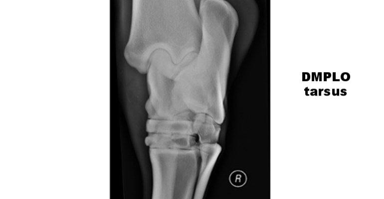
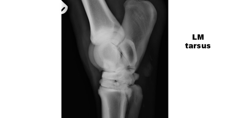
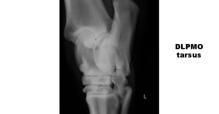
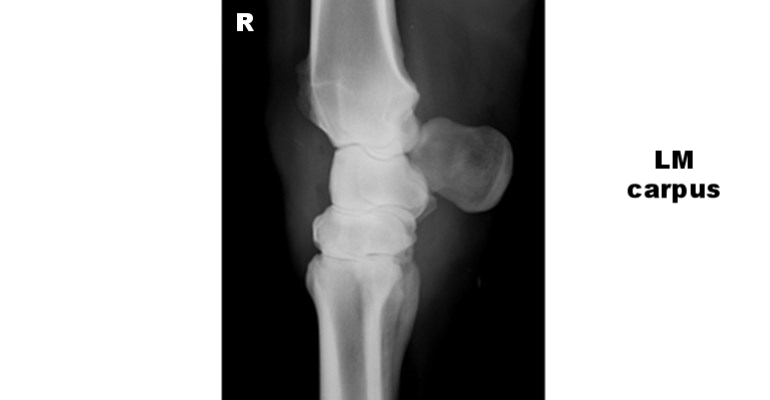
What should my films look like?
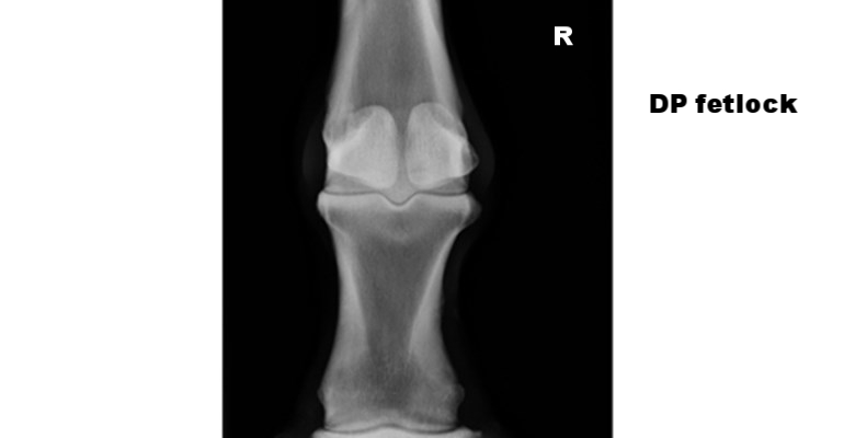
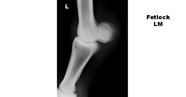
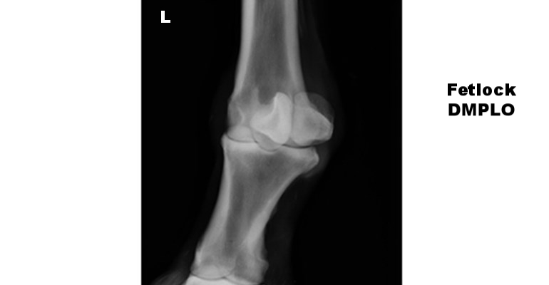
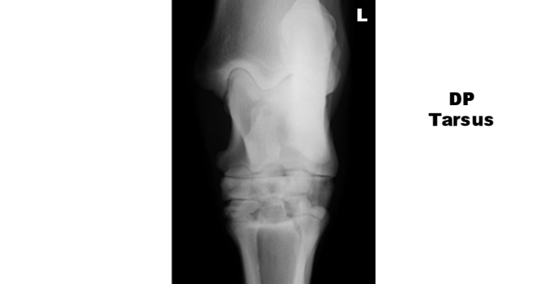
See the image gallery below for examples of some normal LM, DP and lateral/medial oblique views of the carpus ("knee"), tarsus ("hock") and metacarpophalangeal ("fetlock") joints.
For a more detailed guide to radiography of the metacarpophalangeal/metatarsophalangeal ("fetlock") joint please click through to see this guide from BCF Technology.